20th, April, 2025
Ultrasound Case: Ectopic pregnancy
A 28-year-old female with right lower quadrant pain and vaginal spotting
Case Presentation
History
- Chief Complaint: Right lower quadrant pain and vaginal spotting
- History of Present Illness:
- 6 weeks since last menstrual period
- Positive home pregnancy test
- Sharp RLQ pain for 2 days, worsening
- Light vaginal bleeding x 3 days
- No fever or urinary symptoms
- Past Medical History:
- Previous pelvic inflammatory disease (2 years ago)
- No prior pregnancies
- Initial Labs: β-hCG 2,450 mIU/mL, progesterone 8 ng/mL
EMERGENCY FINDING: This patient has classic risk factors (PID history) and symptoms of ectopic pregnancy - requires immediate ultrasound evaluation.
Ultrasound findings
- Extrauterine Gestation sac: Gestation sac containing a fetal pole outside endometrial cavity
- Ring of Fire: Peripheral vascularity around the gestation sac on color Doppler
- Empty Uterus: No Intrauterine pregnancy (IUP)
- Hemoperitoneum: Free fluid in cul-de-sac
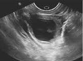
1. Extrauterine sac
- Extrauterine gestation sac containing a fetal pole seen in the right adnexa
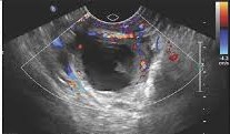
2. Ring of fire
- Periferal vascularity around the extrauterine sac on color doppler application
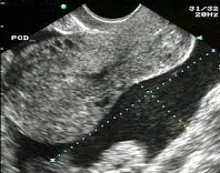
3. Empty uterus and hemoperitoneum in the POD
- No Intrauterine sac and complex free fluid in the posterior cul-de-sac due to hemoperitoneum
Diagnosis: Right adnexal Ectopic pregnancy
Based on history, initial lab results and ultrasound features (Empty uterus, Extrauterine GS, Ring of fire & Hemoperitoneum).
Differentials
- Corpus Luteum Cyst: Adjacent to ovary, thick-walled, no yolk sac/embryo.
- Hemorrhagic Cyst: No peripheral flow, retractile clot, avascular.
- Ovarian Torsion: Absent venous flow, enlarged ovary, no GS.
- Pelvic Inflammatory Disease (PID): Pelvic free fluid, Tubo-ovarian complexes, fluid filled tubes and hypervascularity, bilateral lower abdominal pain, increased yellowish vaginal discharge but negative HCG test.
- Appendicitis: Non-gravid uterus, blind-ending tubular structure.