13th, April, 2025
Ultrasound Case: Thyroiditis
A 42-year-old female with neck swelling, discomfort and fatigue x 3 weeks
Case Presentation
History
- Chief Complaint: Neck discomfort and fatigue x 3 weeks
- History of Present Illness:
- Gradual onset of anterior neck tenderness
- Mild dysphagia but no odynophagia
- Fatigue and weight gain (4kg in 3 weeks)
- No recent fever or viral illness
- Past Medical History: Hypothyroidism (mother and sister)
- Medications: None
Physical Examination
- Neck: Diffusely enlarged thyroid (~2x normal), firm consistency, mild tenderness
- No lymphadenopathy
- No thyromegaly
- Vital Signs: BP 120/80, HR 68, Temp 36.8°C
Initial Labs: TSH 8.2 mIU/L (↑), Free T4 0.8 ng/dL (↓), TPO antibodies >600 IU/mL (↑↑), ESR 22 mm/hr (mildly ↑)
Ultrasound findings
- Parenchymal texture: Markedly heterogeneous with micronodularity (1-6mm)
- Echogenicity: Diffusely hypoechoic compared to strap muscles
- Vascularity: Increased flow with "thyroid inferno" pattern
- Size: Diffuse enlargement (isthmus >5mm)
- Nodules: Several small benign-appearing nodules
- Lymph nodes: Reactive appearing but normal morphology
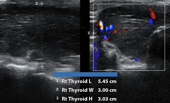
1. Right thyroid lobe
- Diffusely enlarged (5.45cmx3.00cmx3.03cm)
- heterogeneous echotexture
- Diffusely hypoechoic
- Increased flow on color Doppler

2. Left thyroid lobe
- Diffusely enlarged (5.60cmx3.05cmx3.48cm)
- heterogeneous echotexture
- Diffusely hypoechoic
- Increased flow on color Doppler
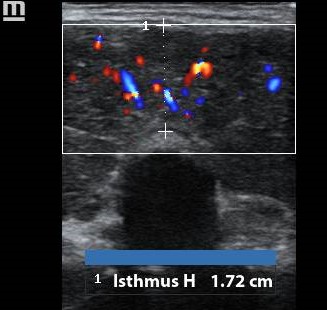
2. Isthmus
- Diffuse enlargement (isthmus = 1.72cm)
Diagnosis: Hashimoto's Thyroiditis (Chronic Lymphocytic Thyroiditis)
Based on characteristic ultrasound findings, elevated TPO antibodies, and clinical presentation.
Differentials
- Subacute Thyroiditis (de Quervain): Painful, recent viral illness, elevated ESR, transient hyperthyroidism. Ultrasound may show focal hypoechoic areas with "washout" vascularity, no micronodularity
- Graves' Disease: Hyperthyroid symptoms, TRAb positive, no tenderness. Ultrasound may show Hypoechoic, hypervascular ("thyroid inferno"), no micronodularity
- Multinodular Goiter: Non-tender, normal thyroid function tests. Ultrasound may show Multiple discrete nodules with normal intervening parenchyma
- Acute Suppurative Thyroiditis: Fever, severe pain, leukocytosis. Ultrasound may show focal hypoechoic area with debris, possible abscess formation