Biliary hamartoma
Introduction
Biliary hamartomas (also known as von Meyenburg complexes) are benign malformations of the biliary tract that appear as small, cystic lesions scattered throughout the liver parenchyma. These developmental anomalies result from failure of remodeling of the ductal plates during embryogenesis.
Clinical presentation
Due to their small sizes, Biliary hamartomas are asymptomatic and usually found incidentally. However, in rare cases, they can potentially cause abdominal pain, jaundice, fever, or portal hypertension if they become large enough to obstruct bile flow or significantly impact liver function
Ultrasound Features
- Multiple small lesions (usually 0.1-1.5 cm in diameter)
- Uniform appearance throughout the liver
- Two morphological patterns:
- Cystic pattern: Anechoic with posterior acoustic enhancement
- Solid-appearing pattern: Hyperechoic or hypoechoic nodules
- No vascular flow on Doppler examination
- No mass effect on surrounding vessels
- Diffuse distribution (though may occasionally cluster in one lobe)
Key Diagnostic Feature: The combination of multiple uniformly sized small cystic/solid lesions distributed throughout the liver parenchyma without vascular flow is highly suggestive of biliary hamartomas.
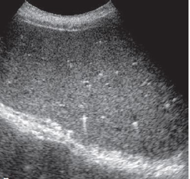
Biliary hamartomas
- Typical ultrasound appearance of multiple small biliary hamartomas
Differential Diagnosis
| Condition | Distinguishing Features |
|---|---|
| Simple hepatic cysts | Larger, variable sizes, solitary or few in number |
| Polycystic liver disease | Numerous larger cysts (>2cm), often with renal cysts |
| Caroli disease | Saccular dilatation of intrahepatic bile ducts, "central dot" sign |
| Metastases | Variable sizes, irregular margins, may show vascularity |
| Microabscesses | Clinical context of infection, may have hypoechoic halo |
Advanced Ultrasound Techniques
Contrast-Enhanced Ultrasound (CEUS)
- No enhancement in cystic lesions
- Possible thin peripheral enhancement in solid-appearing variants
- No late washout (helps differentiate from metastases)
Elastography
- Normal liver stiffness measurements
- Lesions themselves are too small to assess individually
Pitfall: The solid-appearing variant of biliary hamartomas may be mistaken for metastases or other solid lesions. Correlation with other imaging modalities (CT/MRI) or biopsy may be needed in doubtful cases.
Clinical Significance
- Entirely benign with no malignant potential
- Usually asymptomatic (incidental finding)
- No surveillance or follow-up needed when typical features present
- Rare associations:
- Autosomal dominant polycystic kidney disease
- Congenital hepatic fibrosis
Management Note: When characteristic imaging features are present, no further workup is typically required. Biopsy should be avoided unless there is strong suspicion of alternative pathology.
Comparison to other imaging modalities
| Modality | Findings | Advantages |
|---|---|---|
| Ultrasound | Multiple small cystic/solid lesions, no vascularity | First-line, no radiation, real-time evaluation |
| CT | Hypodense lesions, no enhancement | Better for whole-liver evaluation |
| MRI | T2 hyperintense, T1 hypointense, no enhancement | Best for tissue characterization |
References
- Mortelé KJ, Ros PR. Cystic focal liver lesions in the adult: differential CT and MR imaging features. Radiographics. 2001;21(4):895-910.
- Horton KM, Bluemke DA, Hruban RH, et al. CT and MR imaging of benign hepatic and biliary tumors. Radiographics. 1999;19(2):431-51.
- Choi BI, Yeon KM, Kim SH, Han MC. Caroli disease: central dot sign in CT. Radiology. 1990;174(1):161-3.
- Dietrich CF, Mertens JC, Braden B, et al. Contrast-enhanced ultrasound of histologically proven liver hemangiomas. Hepatology. 2007;45(5):1139-45.