Portal hypertension
Introduction
Portal hypertension is a clinical syndrome defined by a pathological increase in the portal venous pressure, with the hepatic venous pressure gradient (HVPG) exceeding 5 mmHg. Ultrasound plays a crucial role in the non-invasive assessment of portal hypertension, providing both direct and indirect signs of this condition.
Clinical Presentation
Patients with portal hypertension may present with:
- Ascites - The most common clinical manifestation
- Gastroesophageal varices - With risk of life-threatening hemorrhage
- Splenomegaly - Due to venous congestion
- Hepatic encephalopathy - From portosystemic shunting
- Caput medusae - Dilated periumbilical veins
Ultrasound findings
Direct Signs
- Portal vein diameter >13 mm (measured at the hepatic hilum during quiet respiration)
- Loss of respiratory phasicity in portal vein Doppler waveform
- Portal vein flow velocity <16 cm/s
- Hepatofugal flow (reversed portal vein flow)
- Portal vein thrombosis (partial or complete)
Indirect Signs
- Splenomegaly (spleen length >12 cm)
- Portosystemic collaterals (varices):
- Paraumbilical vein (recanalized ligamentum teres)
- Coronary-gastric varices
- Splenorenal shunts
- Cavernous transformation of the portal vein (chronic thrombosis)
- Ascites
- Liver parenchymal changes consistent with cirrhosis
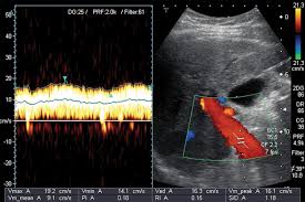
1. Doppler findings
- Doppler ultrasound showing hepatofugal flow in the portal vein
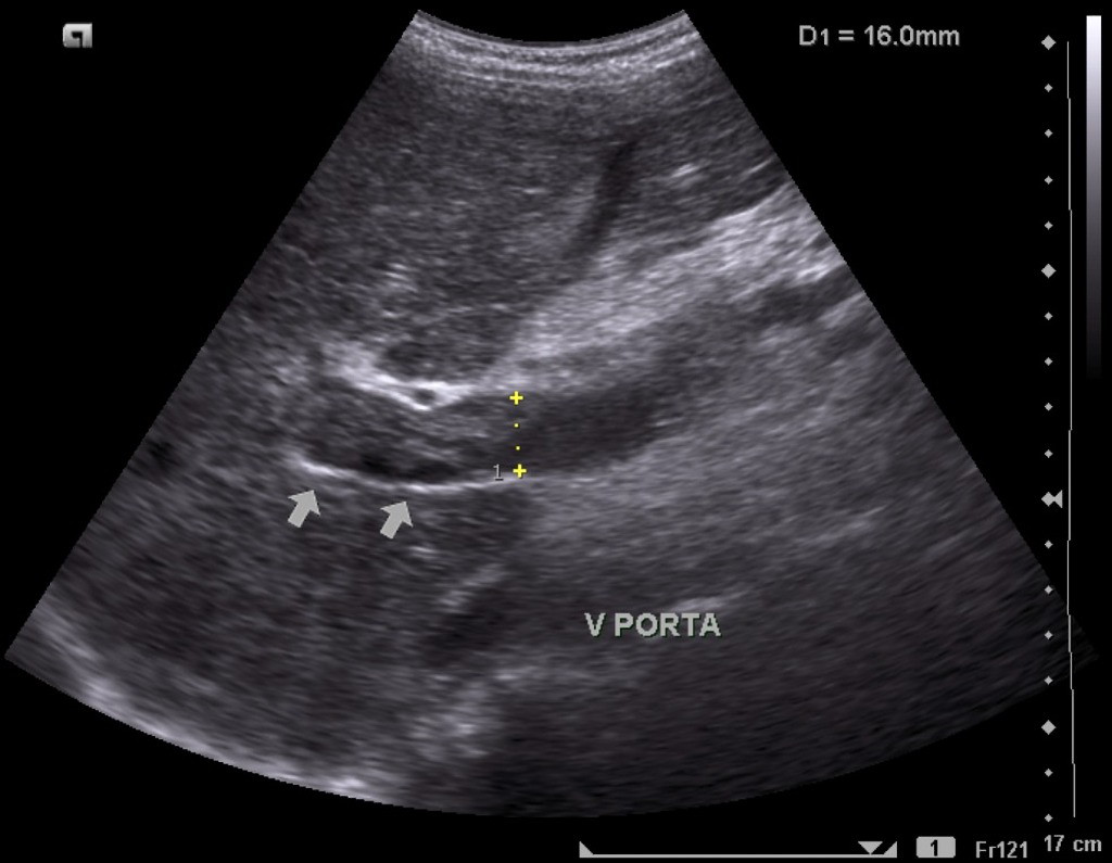
2. Dilatation of portal vein (calipers)
- B-Mode ultrasound showing main portal vein diameter of 15.1 millimeters.
- This is an indirect finding of portal hypertension.
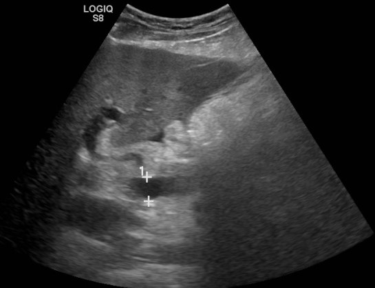
2. Dilatation of splenic vein (calipers)
- Splenic vein dilation, collaterals at the splenic hilum, and Gamna-Gandy bodies in the splenic parenchyma.
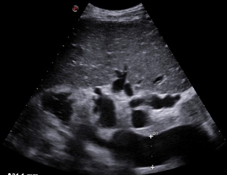
2. Portal vein thrombosis
- Echogenic contents in the portal vein lumen
- Absence of flow on doppler
- Distension of the portal vein and its tributaries
Doppler Ultrasound Parameters
| Parameter | Normal Value | Portal Hypertension |
|---|---|---|
| Portal vein diameter | <13 mm | >13 mm |
| Portal vein velocity | 16-40 cm/s | <16 cm/s |
| Congestion index (Area/Velocity) | <0.07 | >0.07 |
| Flow direction | Hepatopetal | Hepatofugal in advanced cases |
Collateral Pathways
Ultrasound can identify several portosystemic collateral pathways that develop in portal hypertension:
- Gastroesophageal varices - Most clinically significant due to bleeding risk
- Paraumbilical vein - Recanalized ligamentum teres, seen as tortuous vein from left portal vein to abdominal wall
- Splenorenal shunts - Between splenic vein and left renal vein
- Hemorrhoidal plexus - Between superior and middle/inferior rectal veins
- Retroperitoneal collaterals - Between mesenteric veins and retroperitoneal veins
Conclusion
Ultrasound is an invaluable tool in the evaluation of portal hypertension, providing both anatomical and hemodynamic information. While it cannot replace direct pressure measurements, it offers a non-invasive method for diagnosis, assessment of complications, and monitoring of disease progression. The combination of B-mode imaging and Doppler evaluation allows comprehensive assessment of the portal venous system and associated collateral circulation.
References
- Berzigotti A, et al. Ultrasound in portal hypertension - part 1. Ultraschall Med. 2021;42(1):34-57.
- Dietrich CF, et al. Ultrasound in portal hypertension - part 2. Ultraschall Med. 2021;42(2):128-161.
- European Association for the Study of the Liver. EASL Clinical Practice Guidelines for the management of patients with portal hypertension. J Hepatol. 2018;69(4):716-735.
- American College of Radiology. ACR Appropriateness Criteria® for Radiologic Management of Portal Hypertension.