Splenomegaly
Introduction
There are no absolute criteria for the spleen size on ultrasound. When normal, its a little larger than the left kidney. The length should not exceed 15cm in the major axis (from pole to pole). A chronically enlarged spleen may often distort and displace the left kidney, narrowing it in the antero-posterior diameter and width
Clinical presentation
Patients with splenomegaly can present with a variety of symptoms and signs, depending on the underlying cause. The clinical presentation can range from asymptomatic (discovered incidentally) to symptomatic with complications such as Hypersplenism or splenic infarction
Common symptoms
- Left upper quadrant pain or fullness
- Early satiety or abdominal distension. Due to compression of the stomach by the enlarged spleen
- Systemic Symptoms (if due to underlying disease). These include; Fever, Fatigue, recurrent infections and bleeding or bruisng
- Symptoms of complication. Eg Hypersplenism and splenic rapture
Ultrasound features
General Features
- Rounded contour (loss of normal wedge shape)
- Extension below the left costal margin
- Medial displacement of the left kidney
- Compression of adjacent structures (stomach, pancreas)
Doppler Evaluation
- Splenic vein dilation (>10 mm) suggests portal hypertension.
- Increased arterial flow may indicate infection or malignancy.
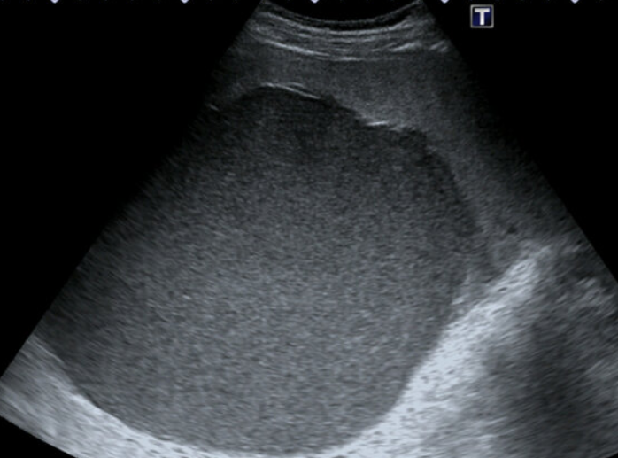
Homogenous splenomegaly
This may be due to:
- Tropical splenomegaly, which includes idiopathic splenomegaly, malaria, trypanosomiasis, leishmaniasis and schistosomiasis
- Sicke cell disease (unless infarcted)
- Portal hypertension
- Leukaemia
- Metabolic disease
- Lymphoma (may contain hyperechoic masses).
- Infections such as rubella and mononucleosis
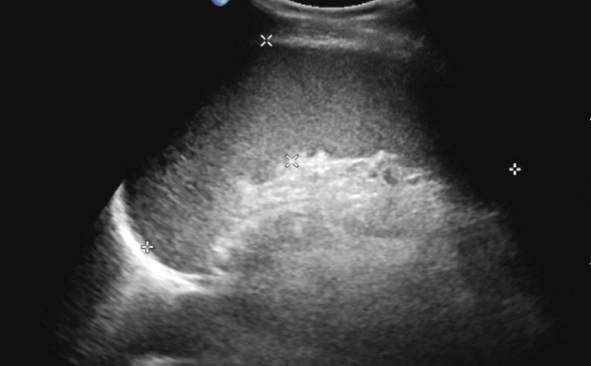
Homogeneous splenomegaly
Note the runded borders of the spleen
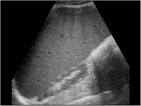
Homogeneous splenomegaly
Enlarged spleen compressing the left kidney
Non-homogenous splenomegaly
This is due to focal abnormalities:
-
Splenic cysts; Anechoic, thin-walled, posterior enhancement
- Congenital cysts
- Pseudocysts
- Hydatid cysts
- Hemangioma; Hyperechoic, well-defined
- Abscess; Hypoechoic, thick-walled, debris
- Lymphoma; Hypoechoic, round masses
- Metastases; Variable (melanoma often hyperechoic)
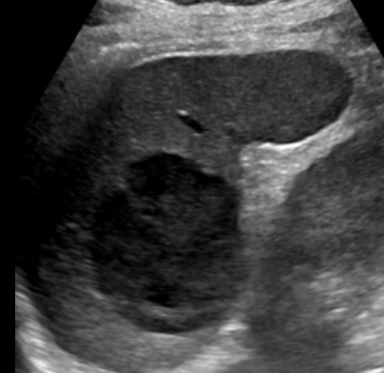
Splenic cyst
- Well-defined hypoechoic intrasplenic cyst
- Low level homogenous echoes (due to deposition of cholesterol crystals)
Splenic abscess
Hypoechoic, thick-walled cystic splenic mass with debris in a patient with Fever and sepsis
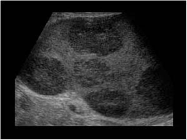
Lymphoma
Multiple hypoechoic, round masses
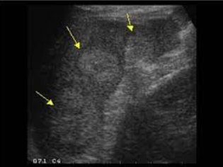
Splenic metastasis
Hyperechoic splenic masses (arrows) in a patient with breast cancer consistent with splenic metastasis
Warning: In trauma patients, splenomegaly increases rupture risk. Assess for subcapsular hematoma or free fluid.
References
- Rumack CM, et al. Diagnostic Ultrasound. 5th ed. Elsevier; 2018.
- Gorg C, et al. Ultrasound of the spleen. Insights Imaging. 2020;11(1):100.
- Kessler N, et al. Evaluation of the spleen with ultrasound. J Radiol. 2004;85(4):655-63.
- Paterson A, et al. Splenic imaging in children. Radiographics. 2009;29(5):1465-85.