17th, April, 2025
Ultrasound Case: Acute splenic sequestration
An 8-year-old male with HbSS disease complaining of left upper quadrant pain
Case Presentation
History
- Chief Complaint: Left upper quadrant pain and fatigue
- History of Present Illness:
- 2-day history of worsening abdominal pain
- Pale appearance noted by parents
- No fever or trauma
- Last transfusion 6 weeks ago
Physical Examination
- Tender splenomegaly (3cm below costal margin)
- Pale conjunctivae
- Hemoglobin: 5.2 g/dL (baseline 8.0)
Clinical Suspicion: Acute splenic sequestration crisis given acute drop in hemoglobin with splenomegaly in a pediatric sickle cell patient.
Ultrasound findings
- Spleen size: Markedly enlarged
- Echotexture: Heterogeneous with hypoechoic areas
- Doppler Flow: Decreased parenchymal flow
- Perisplenic fluid: Small anechoic rim around the spleen
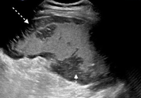
Splenic Sequestration
- Heterogeneous parenchymal echotexture
- Patchy hypoechoic areas
2. Perisplenic fluid
- Anechoic rim around the spleen
Diagnosis: Acute splenic sequestration
Based on clinical findings (Sudden, painful splenic enlargement and signs of acute anemia e.g pallor and fatigue) and ultrasound features (Splenomegaly, parenchymal heterogeneity, decreased venous outflow and patchy hypoechoic areas).
Differentials
- Splenic infarction: Wedge-shaped hypoechoic lesions, no significant enlargement
- Splenic abscess: Hypoechoic lesions with debris/septations + fever
- Splenic rapture: Irregular margin, hemoperitoneum, history of trauma