25th, April, 2025
Ultrasound Case: Hepatic abscess
A 22-year-old male with fever (39.1°C) and RUQ pain for 5 days
Case Presentation
History
- Chief Complaint: Fever (39.1°C) and RUQ pain for 5 days
- History of Present Illness:
- Progressive dull RUQ pain
- High-grade fevers with chills
- Anorexia and 3kg weight loss
- No jaundice or diarrhea
-
Past Medical History:
- Known gallstones
- Type 2 diabetes mellitus
- Hypertension
- Laboratory Findings:
- WBC 18.2 x10³/µL (90% neutrophils)
- CRP 156 mg/L
- ALT 85 U/L, AST 78 U/L, ALP 210 U/L
Physical Examination
- Tender hepatomegaly
- Positive Murphy's sign
Ultrasound findings
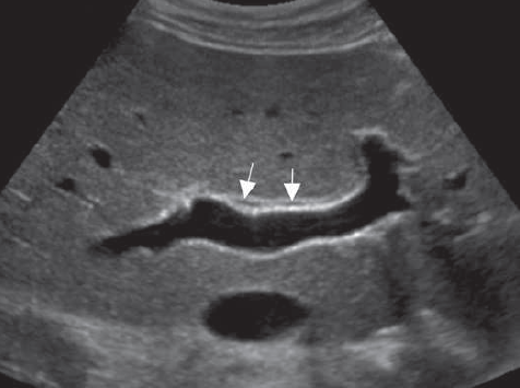
Hypoechoic lesion with irregular walls and internal debris:
- Shape: Ovoid with irregular margins
- Echotexture: Hypoechoic with internal echoes
- Wall characteristics: Thick (4mm), irregular
- Doppler: Hypervascular rim, no internal flow
Hepatic abscess
- Hypoechoic lesion with irregular walls and internal debris
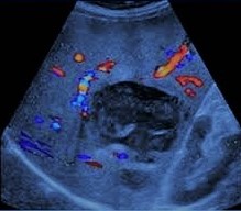
Hepatic abscess
- Periferal hypervascularity
- No internal flow
Diagnosis: Pyogenic liver abscess confirmed by ultrasound-guided aspiration (Klebsiella pneumoniae grown on culture).
Differentials
- Necrotic metastasis: Irregular internal vascularity, known primary.
- Complex hepatic cyst: Thinner walls, no hypervascular rim.
- Echinococcal cyst: Daughter cysts, "water lily" sign.
- Hematoma: Trauma history, evolves over time.