Biliary tree infections
Introduction
Biliary tract infections encompass a wide spectrum of conditions ranging from acute bacterial infections to chronic parasitic infestations. Ultrasound serves as the primary imaging modality due to its excellent soft tissue differentiation, real-time imaging capability, and lack of ionizing radiation. This article details the sonographic appearance of five clinically significant biliary infections:
- Ascending cholangitis - Bacterial infection of obstructed bile ducts
- Liver fluke infections - Parasitic infestations (Clonorchis, Opisthorchis, Fasciola)
- Recurrent pyogenic cholangitis - Oriental cholangiohepatitis
- Biliary ascariasis - Worm infestation of biliary tree
- HIV cholangiopathy - AIDS-related biliary disease
1. Ascending cholangitis
Acute bacterial infection secondary to biliary obstruction, most commonly from stones (70%), followed by strictures and tumors.
Pathophysiology
Obstruction → bacterial overgrowth (E. coli, Klebsiella, Enterococcus) → increased ductal pressure → systemic infection.
Ultrasound Findings
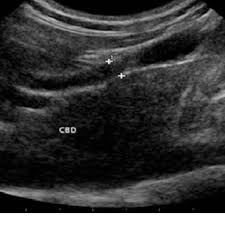
- Bile duct dilation (CBD >8mm, intrahepatic >2mm)
- Echogenic debris within ducts (pus/sludge)
- Pneumobilia (20% of cases) - bright echoes with "dirty" shadowing
- Gallstones or other obstructive lesions
- Portal vein thrombosis in severe cases
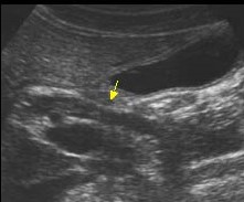
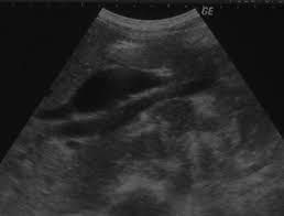
Cholangitis
- Bile duct dilation
- Echogenic debris within the duct
Cholangitis
- Echogenic focui with shadows within the dilated duct
2. Liver Fluke Infections
Chronic parasitic infestations caused by Clonorchis sinensis, Opisthorchis viverrini (Asian liver flukes), and Fasciola hepatica (sheep liver fluke).
Epidemiology & Life Cycle
Endemic in Southeast Asia. Humans ingest metacercariae in raw fish (Clonorchis) or water plants (Fasciola) → larvae migrate to bile ducts → mature into adult flukes.
Ultrasound Findings
- Ductal wall thickening (fibrosis from chronic irritation)
- Intrahepatic duct dilation with normal CBD (60% of cases)
- Fluke visualization (2-10mm echogenic foci in ducts)
- Periductal fibrosis (hyperechoic streaks)
- Gallbladder sludge/stones (common complication)
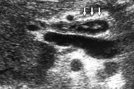
Liver fluke
- Echogenic foci in the dilated intrahepatic duct
Liver fluke
- Intrahepatic duct dilation
- Periductal fibrosis
3. Recurrent Pyogenic Cholangitis (RPC)
Also called Oriental cholangiohepatitis, common in Southeast Asia. Characterized by recurrent bacterial cholangitis with intrahepatic pigment stones.
Ultrasound Findings
- Segmental intrahepatic duct dilation (left lobe predominance)
- Intraductal stones (cast-like, non-shadowing)
- Ductal strictures with abrupt caliber changes
- Parenchymal atrophy of affected segments
- Portal vein thrombosis in advanced cases
Pathogenesis
Chronic parasitic infection (flukes/ascaris) → bile stasis → bacterial colonization → stone formation → recurrent infection cycles.
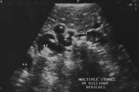
Reccurent pyogenic cholangitis
- Segmental intrahepatic duct dilatation
- Intraductal stones
Reccurent pyogenic cholangitis
- Segmental intrahepatic duct dilatation
4. Biliary Ascariasis
Infestation by Ascaris lumbricoides, the largest intestinal nematode (15-35cm long).
Ultrasound Findings
- Long tubular structure in bile ducts (4-6mm diameter)
- Parallel echogenic lines (worm's digestive tract)
- Spaghetti sign - coiled worms in gallbladder
- Real-time movement of worms (diagnostic)
- Ductal dilation proximal to obstruction
Clinical Management
Albendazole/mebendazole therapy. ERCP for extraction if worms don't retreat to intestine within 3 days.
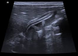
Biliary ascariasis
- Long tubular structure in the bile duct
- Parallel echogenic lines
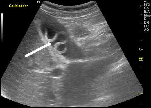
Biliary ascariasis
- Spaghetti sign (Coiled worm in the gallbladder lumen)
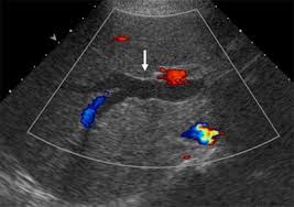
5. HIV Cholangiopathy
AIDS-related biliary disease occurring when CD4 counts <100 cells/mm³, caused by opportunistic infections (Cryptosporidium, CMV, Microsporidia).
Ultrasound Findings
- Ductal wall thickening (>1.5mm)
- Strictures (focal or diffuse)
- Papillary stenosis (CBD dilation >12mm)
- Intrahepatic duct irregularity (beaded appearance)
- Gallbladder wall thickening (acalculous cholecystitis)
Treatment Approach
Antiretroviral therapy (ART) is primary treatment. ERCP with sphincterotomy for symptomatic relief.

HIV cholangiopathy
- Scan at the porta hepatis shows tapered narrowing of a dilated distal common bile duct (curved arrows) image and wall thickening (straight arrow)
HIV cholangiopathy
- Papillary stenosis
Comparative Ultrasound Features
| Condition | Key Ultrasound Feature | Pathognomonic Sign |
|---|---|---|
| Ascending cholangitis | Dilated ducts + echogenic debris | Pneumobilia |
| Liver flukes | Ductal wall thickening | Visible flukes in ducts |
| RPC | Left lobe ductal dilation | Cast-like intraductal stones |
| Ascariasis | Long tubular structure | Parallel echogenic lines |
| HIV cholangiopathy | Ductal wall thickening | Beaded intrahepatic ducts |
References
- Lim JH. Liver flukes: the malady neglected. Korean J Radiol. 2011;12(3):269-279. doi:10.3348/kjr.2011.12.3.269
- Kiriyama S, Kozaka K, Takada T, et al. Tokyo Guidelines 2018: diagnostic criteria and severity grading of acute cholangitis. J Hepatobiliary Pancreat Sci. 2018;25(1):17-30. doi:10.1002/jhbp.512
- Chan HH, Lai KH, Lin CK, et al. Endoscopic diagnosis and management of biliary parasitosis. Diagn Ther Endosc. 2011;2011:376023. doi:10.1155/2011/376023
- Lim JH, Kim SY, Park CM. Parasitic diseases of the biliary tract. AJR Am J Roentgenol. 2007;188(6):1596-1603. doi:10.2214/AJR.06.1172
- Catalano OA, Sahani DV, Forcione DG, et al. Biliary infections: spectrum of imaging findings and management. Radiographics. 2009;29(7):2059-2080. doi:10.1148/rg.297095051
- Raman SP, Horton KM, Fishman EK. Multimodality imaging of pancreatic and biliary neoplasms. Surg Oncol Clin N Am. 2014;23(4):649-689. doi:10.1016/j.soc.2014.06.002
- Baron TH, Davee T. AIDS cholangiopathy. Curr Treat Options Gastroenterol. 2003;6(2):111-117. doi:10.1007/s11938-003-0002-0
- Park MS, Yu JS, Kim KW, et al. Recurrent pyogenic cholangitis: comparison between MR cholangiography and direct cholangiography. Radiology. 2001;220(3):677-682. doi:10.1148/radiol.2203001424
- Rumack CM, Levine D. Diagnostic Ultrasound. 5th ed. Elsevier; 2018.
- American Institute of Ultrasound in Medicine (AIUM). AIUM practice guideline for the performance of ultrasound examinations of the abdomen and/or retroperitoneum. J Ultrasound Med. 2021;40(7):E1-E16. doi:10.1002/jum.15607