Polycystic Liver Disease
Introduction
Polycystic liver disease (PLD) is a genetic disorder characterized by the presence of multiple cysts scattered throughout the liver parenchyma. While often associated with autosomal dominant polycystic kidney disease (ADPKD), PLD can also occur as an isolated condition (autosomal dominant polycystic liver disease, ADPLD). Ultrasound plays a crucial role in diagnosis, monitoring, and management of this condition.
Ultrasound Features
Characteristic Ultrasound Features
- Multiple cysts: Typically >20 cysts distributed throughout both liver lobes
- Cyst morphology: Round or oval anechoic structures with sharp margins
- Cyst size: Varies from few millimeters to several centimeters
- Cyst walls: Thin, smooth walls (<1mm thickness)
- Acoustic enhancement: Posterior acoustic enhancement behind cysts
- No internal flow: Absence of internal vascularity on Doppler
- Parenchymal changes: Normal liver parenchyma between cysts in early stages
Advanced Disease Features
In advanced PLD, ultrasound may demonstrate:
- Massive hepatomegaly with abdominal distension
- Near-complete replacement of liver parenchyma by cysts
- Compression of adjacent structures (IVC, portal vein, bile ducts)
- Complicated cysts (hemorrhage, infection, rupture)
- Signs of portal hypertension in severe cases
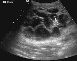
1. Classic ultrasound appearance
- Classic ultrasound appearance of polycystic liver disease showing numerous cysts of varying sizes
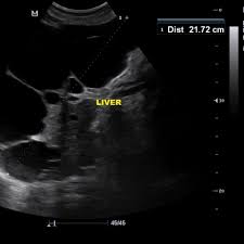
2. Advanced Disease Features
- Advanced PLD with near-total parenchymal replacement and mass effect
Diagnostic Criteria
Ultrasound Diagnostic Criteria for PLD
For ADPKD-associated PLD:
- Age <40 years: ≥3 liver cysts
- Age 40-59 years: ≥4 liver cysts
- Age ≥60 years: ≥5 liver cysts
For isolated ADPLD:
- Age <40 years: ≥4 liver cysts
- Age ≥40 years: ≥10 liver cysts
- No renal cysts (or very few)
- Family history of PLD
Grading System for Disease Severity
| Grade | Description | Ultrasound Findings |
|---|---|---|
| I (Mild) | <10 cysts | Normal liver architecture, cysts <5cm |
| II (Moderate) | 10-20 cysts | Mild parenchymal replacement, some cysts >5cm |
| III (Severe) | >20 cysts | Significant parenchymal replacement (>50%) |
| IV (Massive) | Innumerable cysts | Near-total parenchymal replacement, hepatomegaly |
Differential Diagnosis
| Condition | Distinguishing Features | Key Differences from PLD |
|---|---|---|
| Simple hepatic cysts | Solitary or few cysts | Fewer in number, no genetic basis |
| Biliary hamartomas | Multiple small cysts (<1.5cm) | Uniform small size, no family history |
| Caroli disease | Cystic biliary dilatation | Communicates with biliary tree, central dot sign |
| Hydatid cysts | Complex cystic lesions | Daughter cysts, wall calcifications |
| Metastatic disease | Complex cystic masses | Thick walls, solid components, known primary |
Clinical Implications
Complications Detectable by Ultrasound
- Cyst hemorrhage: Internal echoes, fluid-debris levels
- Cyst infection: Thickened walls, internal debris, gas bubbles
- Biliary obstruction: Dilated intrahepatic bile ducts
- Vascular compression: Narrowed hepatic veins/IVC, portal vein
- Cyst rupture: Free fluid, irregular cyst margins
Monitoring Recommendations
- Baseline ultrasound for all ADPKD patients
- Annual ultrasound for symptomatic patients or Grade III/IV disease
- Every 2-3 years for asymptomatic Grade I/II disease
- Immediate ultrasound for acute symptoms (pain, fever, jaundice)
- Preoperative mapping for planned interventions
References
- Gevers TJ, Drenth JP. Diagnosis and management of polycystic liver disease. Nat Rev Gastroenterol Hepatol. 2013;10(2):101-108.
- Van Aerts RMM, Van de Laarschot LFM, Banales JM, Drenth JPH. Clinical management of polycystic liver disease. J Hepatol. 2018;68(4):827-837.
- Hoevenaren IA, Wester R, Schrier RW, et al. Polycystic liver: clinical characteristics of patients with isolated polycystic liver disease compared with patients with polycystic liver and autosomal dominant polycystic kidney disease. Liver Int. 2008;28(2):264-270.
- Qian Q, Li A, King BF, et al. Clinical profile of autosomal dominant polycystic liver disease. Hepatology. 2003;37(1):164-171.
- Mavilia MG, Pakala T, Molina M, Wu GY. Differentiating Cystic Liver Lesions: A Review of Imaging Modalities, Diagnosis and Management. J Clin Transl Hepatol. 2018;6(2):208-216.
- European Association for the Study of the Liver. EASL Clinical Practice Guidelines on the management of cystic liver diseases. J Hepatol. 2022;77(4):1083-1108.