24th, April, 2025
Ultrasound Case: Cholangitis
A 72-year-old male with fever, jaundice, and RUQ pain (Charcot's triad)
Case Presentation
History
- Chief Complaint: Fever, jaundice, and RUQ pain (Charcot's triad)
- Duration: Symptoms progressing over 48 hours
- History of Present Illness:
- Colicky RUQ pain radiating to back
- High-grade fever (39.2°C) with chills
- Progressive yellowish discoloration of skin
- Dark urine and pale stools
-
Past Medical History:
- Known gallstones
- Type 2 diabetes mellitus
- Hypertension
- Medications: Metformin, lisinopril
-
Laboratory Findings:
- WBC 19.5 x10³/µL with 90% neutrophils
- Total bilirubin 5.2 mg/dL (direct 4.3)
- ALP 520 U/L, GGT 380 U/L
- AST 180 U/L, ALT 160 U/L
Physical Examination
- Vital Signs: Temp 39.1°C, HR 112, BP 90/60, RR 22
- General: Icteric sclerae, dry mucous membranes
- Abdomen:
- Tenderness in RUQ with guarding
- Positive Murphy's sign
- No rebound tenderness
- No palpable masses
- Neurologic: Mild confusion (new onset)
Clinical Suspicion: Acute ascending cholangitis with sepsis (Reynolds' pentad present) secondary to likely choledocholithiasis given history of gallstones and characteristic clinical presentation. Urgent ultrasound needed to evaluate for biliary obstruction and guide emergent decompression.
Ultrasound findings
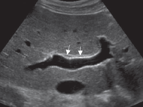
- Bile duct dilation:
- Common bile duct diameter: 12mm
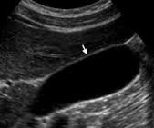
- Intrahepatic ducts >2mm diameter
- "Too many tubes" sign at porta hepatis
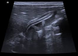
- Obstructing lesion:
- 9mm hyperechoic stone in distal CBD
- Posterior acoustic shadowing
- Proximal ductal dilation
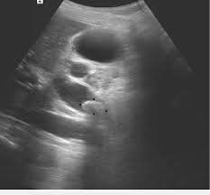
- Gallbladder findings:
- Multiple small gallstones
- Wall thickness: 4mm (mild edema)
CBD dilation and choledocholithiasis
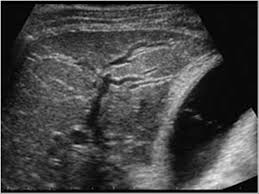
Intrahepatic duct dilation

Multiple gallstones
Diagnosis: Acute obstructive cholangitis secondary to choledocholithiasis, confirmed by ultrasound findings of biliary obstruction with ductal dilation and visualized CBD stone.
Differentials
- Acute cholecystitis: GB wall thickening (>3mm), Pericholecystic fluid, No bile duct dilation.
- Viral hepatitis: Diffuse liver heterogeneity, No ductal dilation, Normal GB wall.
- Pancreatitis: Pancreatic edema, Peripancreatic fluid, May have secondary CBD dilation.
- Biliary stricture: Focal duct narrowing, Gradual symptom onset, No fever typically.
- Cholangiocarcinoma: Mass lesion at duct, Abrupt duct cutoff, No visible stones.